 ×
×
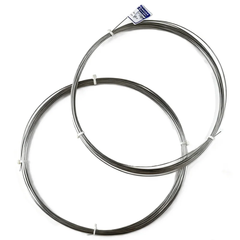
In the world of modern medicine, few materials have had as profound an impact as Nitinol, a nearly equiatomic alloy of nickel and titanium. Since its discovery in the 1960s, Nitinol has evolved from a laboratory curiosity into a cornerstone of minimally invasive surgery, interventional radiology, and implantable device technology. Its two extraordinary properties—the shape memory effect and superelasticity—allow medical devices to do what no conventional metal can: compress into a tiny form for delivery, then autonomously expand into a precisely designed shape inside the human body. Today, Nitinol is found in millions of medical devices, from lifesaving cardiovascular stents to orthodontic wires that gently move teeth.
Before exploring its applications, it is essential to understand the material characteristics that make Nitinol so valuable in a biological environment.
Superelasticity allows Nitinol to undergo large deformations (up to 8–10% strain) and instantly recover its original shape upon unloading. For a medical device, this means a guide wire can be bent around tortuous cerebral vessels without kinking, or a stent can be crimped onto a delivery catheter and later spring open without permanent deformation.
The shape memory effect enables devices to be “programmed” with a specific shape at a high temperature. After cooling, they can be deformed into a compact form. When warmed to body temperature (37 °C), they return to the programmed shape, generating a gentle but continuous force. This property is ideal for selfexpanding implants that deploy precisely when they reach body temperature.
Biocompatibility is another critical factor. Nitinol forms a stable, protective titanium dioxide (TiO₂) layer on its surface, which resists corrosion in the harsh environment of blood and tissue. Extensive clinical use has confirmed its longterm safety, though careful processing is required to minimize nickel ion release.
Radiolucency and MRI compatibility are added benefits. Nitinol is less radiopaque than stainless steel or cobaltchromium, but it can be combined with radiopaque markers. It is also nonferromagnetic, making it safe for magnetic resonance imaging (MRI).
The cardiovascular system was the first major clinical arena for Nitinol. The alloy’s flexibility and selfexpansion properties revolutionized the treatment of arterial blockages and structural heart disease.
Unlike coronary stents (which are typically balloonexpandable stainless steel or cobaltchromium), peripheral arteries—such as the femoral, iliac, and carotid arteries—are subject to bending, torsion, and compression. Nitinol stents, with their superelasticity, maintain patency under these dynamic forces. A Nitinol stent is crimped onto a delivery catheter, inserted through a small incision, and positioned under fluoroscopy. Once released, it expands to its predetermined diameter and provides radial strength to keep the vessel open. The selfexpansion also reduces the risk of vessel rupture compared to balloonexpanded devices.
In treating abdominal aortic aneurysms, large Nitinolbased stentgrafts are used to exclude the aneurysm sac from circulation. The selfexpanding Nitinol framework anchors the graft fabric to the healthy vessel wall above and below the aneurysm. Because Nitinol can be collapsed into a relatively lowprofile delivery system, these complex devices can be inserted through the femoral artery, avoiding open abdominal surgery.
The transcatheter aortic valve replacement (TAVR) revolution relies heavily on Nitinol. The valve prosthesis consists of a Nitinol frame that holds a bioprosthetic leaflet. The frame is compressed into a delivery catheter, advanced to the heart, and expanded to replace a diseased aortic valve. Nitinol provides the precise balance of radial force and conformability needed to anchor the valve without damaging surrounding structures.
Nitinol is also used in occluder devices (such as those for patent foramen ovale and atrial septal defects), embolic protection filters (captured during carotid stenting), and retrievable vena cava filters (designed to trap blood clots). In all these applications, the alloy’s ability to collapse for delivery and expand upon deployment is indispensable.
The musculoskeletal environment poses unique challenges: high cyclic loads, variable anatomy, and the need for secure fixation. Nitinol has found a niche in specialized orthopedic implants.
Spinal spacers and fusion devices made from Nitinol can be inserted through a small incision and then expanded to restore disc height. This minimally invasive approach reduces muscle damage and accelerates recovery compared to traditional open spinal fusion.
Bone anchors and staples using the shape memory effect provide compression across fractures or osteotomies. A Nitinol staple is cooled, spread apart, inserted into predrilled holes, and then warmed by body heat. As it returns to its original shape, it compresses the bone fragments together—a concept known as “memory compression.” This technique is used in foot and hand surgery, as well as in joint fusion procedures.
Scoliosis correction rods made of Nitinol offer dynamic stabilization. Unlike rigid stainless steel rods, superelastic Nitinol rods allow controlled motion while maintaining correction, potentially reducing the risk of adjacent segment disease.
Orthodontics was one of the earliest adopters of Nitinol. Orthodontic archwires made from superelastic Nitinol apply a constant, light force to move teeth, even as the teeth shift. This is a dramatic improvement over stainless steel wires, which lose force rapidly and require frequent tightening. The result is more efficient tooth movement, reduced patient discomfort, and fewer office visits.
Beyond archwires, Nitinol is used in endodontic files for root canal treatment. Superelastic files can navigate the curved canals of teeth with less risk of breakage, improving the success rate of the procedure. Additionally, shapememory NiTi files can be designed to adapt to the canal anatomy.
The superelasticity of Nitinol has enabled the development of instruments that can pass through narrow channels and then deploy complex tools at the target site.
Atrial septal defect closure devices and left atrial appendage occluders rely on Nitinol frames that expand to fit the anatomy.
Basket retrievers for kidney stones and clot retrieval devices for stroke (mechanical thrombectomy) use Nitinol to create expandable nets that capture stones or clots. The devices are delivered through microcatheters and then open like a cage.
Laparoscopic instruments with Nitinol components offer enhanced flexibility and the ability to articulate within the abdominal cavity without sacrificing strength.
In many of these tools, the “memory” of Nitinol allows the device to be folded into a delivery sheath and later assume a complex threedimensional shape that matches the anatomy.
Despite its remarkable advantages, Nitinol presents specific challenges for medical device design and manufacturing.
Nickel hypersensitivity is a concern for a small percentage of patients. While the stable titanium oxide layer minimizes nickel release, some individuals may still experience allergic reactions. Surface treatments and coatings are being developed to further reduce nickel exposure.
Fatigue resistance is critical for implants that undergo millions of cycles (e.g., heart valves, stents). The fatigue behavior of Nitinol is complex and depends on processing, surface quality, and stress levels. Manufacturers must rigorously test devices to ensure longterm durability.
Fabrication complexity makes Nitinol difficult to machine, weld, and join. Laser cutting of Nitinol tubing is the dominant manufacturing method for stents, but heataffected zones can alter transformation properties. Precise thermal processing is essential to achieve the desired transition temperatures.
Radiopacity is inherently lower than that of stainless steel or platinumiridium, so many devices incorporate radiopaque markers (e.g., tantalum or gold) to aid visualization during implantation.
The versatility of Nitinol continues to drive innovation. Several emerging directions promise to expand its medical impact.
Additive manufacturing (3D printing) of Nitinol is being explored to create patientspecific implants with complex geometries that cannot be achieved by traditional machining. Customized bone fixation devices, porous scaffolds for tissue engineering, and personalized stents are active research areas.
Biodegradable Nitinol is an area of investigation. By controlling composition and processing, researchers aim to create implants that provide temporary support and then gradually degrade or be absorbed, eliminating the need for removal surgery.
Sensors and smart implants that use the electrical resistance change associated with phase transformation could allow Nitinol implants to double as sensors, reporting load, temperature, or deformation wirelessly.
Combination devices that integrate drug delivery with Nitinol structures are already in clinical use (e.g., drugeluting stents with Nitinol platforms). Future iterations may incorporate bioactive coatings or local drug reservoirs to further improve outcomes.
Nitinol has fundamentally changed the practice of minimally invasive medicine. Its ability to be compressed, delivered through tiny incisions, and then reexpand into a perfectly fitting implant has made procedures safer, reduced recovery times, and expanded treatment options for patients who were once considered too highrisk for surgery. From the beating heart to the curved canals of a tooth, Nitinol’s unique properties—superelasticity, shape memory, and biocompatibility—have enabled devices that act like living tissues: flexible, resilient, and perfectly adapted to their environment. As manufacturing techniques advance and our understanding of the material deepens, Nitinol will undoubtedly continue to shape the future of medical technology, one “remembered” shape at a time.
Copyright © 2026 Shenzhen Starspring Materials.,Ltd. All rights reserved. - Privacy policy


 Hot News